Written by Taylor Campbell
6. 2. 26
It's only a Failed Experiment if no one Sees it.

It’s a tale as old as time (or at least as old as the 16th century): The failed experiment.
You ran an experiment that didn’t work. So you adjusted. And you ran it again. And again. And again. And again. Until, finally…
She had to be laid to rest.
Not all experiments are made to see the light of day in a journal. But sometimes they are made to be seen somewhere else— somewhere where art and science collide.
IMAGINE SOMETHING DIFFERENT
The cell culture that grew something foreign. The UV lit gel with no DNA bands. The histology stain that never quite worked right. The microscopy image that never made it off the harddrive.
When experiments fail, we think about the time spent and the money wasted. We think about evolving and moving forward. We think about moving on—about facing the next problem.
But we forget about the art left behind.
Scientists produce gorgeous images in the lab every day. These images exist almost as a byproduct of scientific discovery—a necessary step along the way to the final data analysis and publication. By viewing these images as art, we give them permission to exist beyond their scientific purpose. They become more than data, transforming into stories, patterns, windows into other worlds. This perspective shift transforms science communication, removing the barrier for people who may never read a scientific journal article, sparking people to engage with science through wonder first and understanding second.
Thinking about scientific images as abstract art allows us to take what we know and imagine something different.
Like a failed PCR experiment-turned-artist collage.
(but first a quick and, hopefully, painless science lesson to get us all on the same page)
WHERE DOES THIS STEM-STRACT ART COME FROM?
Microscopy images
Using a microscope to study tissues, organisms, or other small structures too small to be seen with the naked eye.
Gel electrophoresis
Method for separating and visualizing biological molecules, like DNA, by size using a porous gel matrix and an electric current.
Protein crystallography
Technique used to determine the three-dimensional structure of proteins.
Culture plates
A shallow, flat-bottomed container, called a petri dish, used in microbiology to grow and maintain microorganisms like bacteria and fungi.
Fluorescence microscopy
Microscopy technique using fluorescent light emitted by a specimen to generate high-contrast images of structures or molecules.

QUICK FOR THE PEOPLE IN THE BACK: A SCIENCE LESSON DETOUR
FINDING A DNA NEEDLE IN A LIBRARY HAYSTACK
Think about DNA like a public library—three floors of countless genres. You’re looking for one specific sentence hidden somewhere in thousands of books.
PCR (polymerase chain reaction) is like having a (very advanced) photocopier that can find that sentence and make millions of copies of it. This technique is used because a sample may only contain a tiny amount of DNA—not enough to study—but after PCR, there are enough copies to detect, analyze, or sequence.
After PCR, you want to check whether you copied the right sentence (or that you actually copied something at all). This is where our gel comes in.
GELS: NOT JUST AN EARLY 2000s HAIR PRODUCT
Imagine a tray of really thick Jell-O with tiny holes cut out of one end. Now imagine dropping PCR samples into those holes and turning on an electric current. The current will move the DNA through the jelly matrix toward the opposite end.
Gel electrophoresis separates DNA fragments by size, moving them through the gel in bands: Small DNA fragments move quickly, while large fragments move more slowly. Gels use a special dye that causes these DNA fragments to show up under UV light.
Since we know how long our target sentence was, we can see if the size of the band on the UV-lit gel matches the size of our expected target. If it matches up, we can be confident that our PCR photocopier accurately detected and copied our target.
Of course, that’s assuming we get a DNA band at all. For our PCR photocopier to copy our target, we have to tell it exactly what we’re looking for. We do this by designing primers—these primers search the library of books and latch onto our specific sentence, causing copies to only be created for this one section.

AND NOW, BACK TO THE SHOW
In PCR, you use primers to copy a target segment of DNA to measure the amount in a sample. Easy enough right?
Nice try.
For PCR to work, your primers have to successfully latch onto your target. And that’s the tricky part—primers don’t always like to work, and when they do, they only work efficiently at very specific temperature ranges (like me). You can test if your primers worked by running a gel— if your primers successfully copied your target, you should see a gorgeous little band appear (there’s a little more to it, but that’s the important bit). And when your primers don’t work… you see nothing but the dyes used in the assay.
If you ever find yourself in the oh so fun position of troubleshooting primers, get comfortable— you’ll probably be there for a while. But you’ll get really comfortable running gels.

FLIPPING FAILURE: SCIENTISTS TO ARTISTS
Failure is a hallmark of good science—it leads us on a path of creative solutions and perseverance.
Most experiments don’t work the first time, and some won’t ever work at all. Being comfortable with failure is a necessary part of science, but that doesn’t mean we don’t have an opportunity to flip these failures into something new.
Something inspiring. Something that gives science a new purpose. Something that shows:
Scientists can be creative too.
Take a gel collage, for instance.
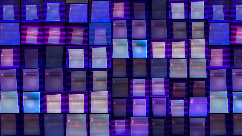

Created by Oviyanna Umoh, this STEM-stract gel collage captures the beauty hidden within scientific setbacks. While spending an entire summer troubleshooting PCR primers, Oviyanna photographed each unsuccessful gel before it was discarded. What emerged was not a record of failure, but a visual diary of persistence—each gel representing another step toward discovery.
Our platform, Coffee Table Science, gives images from failed experiments a new way to live. These images don’t lack intrigue or beauty—they lack statistical significance, and that’s not a good enough reason for them to be tucked away.
We are transforming what most scientists view as failure into something entirely different: a visual testament to resilience and the often-unseen creativity in the scientific process.
Dare to imagine something different and join us where art and science collide.